
Core facilities provide expert training, scientific resources and equipment that might not otherwise be affordable or accessible. The BRI offers a range of core facilities to ensure that BRI members have access to the most advanced technologies for neuroscience research.
Electron Microscopy Core Facility
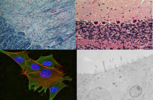
The Electron Microscopy Core Facility, in the Center for Health Sciences room 63-377, houses a JEOL 100CX transmission electron microscope. A TMC MTX and Reichert Ultracut ultramicrotome are available for use by trained personnel. (NOTE: The facility does not supply diamond knives; you must use your own). The Core provides service, advice, and training in fixation and embedding of specimens, thin sectioning, and use of the electron microscopes (with or without assistance), gold labeling, negative stain, and light microscopy of plastic embedded materials. The facility offers advice on appropriate preparatory procedures and other technical matters. Training and assistance in the use of the electron microscope is also offered.
For prices, questions, or consultation, please contact Chunni Zhu (Facility Director) at 310-825-9848 or via email at chunnizhu@mednet.ucla.edu.
Microscopic Techniques Laboratory
 The Microscopic Techniques Laboratory is located in the SEMEL Institute for Neuroscience in room 78-177. This facility can assist you in preparing tissue specimens for light microscopic observation and consult with investigators and their staff on best methodology. The Core provides service and training in various histological procedures. These include immunohistochemistry as well as standard stains like H&E or cresyl violet as well as more specialized stains. The Core facility has setups for paraffin, vibratome and cryostat sectioning as well as plastic embedding and sectioning. The laboratory also has a Nikon photomicroscope with a digital camera.
To use the laboratory and its facilities, you must obtain an Investigator Authorization Form from the Microscopic Techniques Laboratory. For prices, questions, or consultation, please contact Chunni Zhu (Facility Director) at 310-825-9848 or via email at chunnizhu@mednet.ucla.edu.
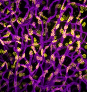
CNSI Imaging Facility
 This facility is run by CNSI and offers many cutting edge light imaging technologies.
For more information, please visit http://alms.cnsi.ucla.edu.
For specific questions please contact Dr. Matt Schibler at: alms@cnsi.ucla.edu.
Neurospecimen Bank
Postmortem Human Frozen Brain Tissue and Matched Cerebrospinal Fluid (CSF) and Blood are Available for Scientists to Search for Etiopathogeneses of Human Disease.
The National Neurological Research Specimen Bank and the Multiple Sclerosis Human Neurospecimen Bank, located at VA West Los Angeles Healthcare Center, maintains a collection of quick frozen and formalin fixed postmortem human brain tissue and frozen cerebrospinal fluid (CSF) from patients with neurological diseases (including Alzheimer’s Disease, amyotrophic lateral sclerosis, depressive disorder/suicide, epilepsy, Huntington’s disease, multiple sclerosis, Parkinson’s Disease, progressive supranuclear palsy, schizophrenia, stroke/CVA and other less common diseases). Full inventory is available upon request. Diagnoses are documented by clinical medical records and gross/microscopic neuropathology.
Special features of the Bank are as follows:
1) Serial digital images of coronal sections (7 mm thick and obtained before quick freezing) are available for selecting samples to be studied.
2) Microscopic neuropathology is available on each dissected sample and the dissected sample’s localization is sketched on the gross coronal section image from which it came.
3) Plaques of demyelination are classified as active, chronic active or inactive, and a shipment includes adjacent normal appearing white and nearby gray matter from the same case (they serve as a type of control).
4) Ice artifact is minimized and it does not interfere with in situ hybridization or in situ PCR or immunocytochemistry.
5) Tissue samples have been used for harvesting enough mRNA for microarray assay plates.
6) CSF cells and cell-free CSF are available pre- and postmortem as is serum, plasma and buffy coats. They are stored quick frozen (full inventory is available upon request).
The Bank is supported by NIH (NINCDS/NIMH), the National Multiple Sclerosis Society and Veterans Affairs West Los Angeles Healthcare Center. For further information on tissues/CSF available and how to access them, contact:
Rashed M. Nagra, Ph.D.
Director/Principal Investigator, Brain Bank
Neurology Research (127A)
VA West Los Angeles Healthcare Center
11301 Wilshire Blvd, Los Angeles, CA 90073
(310) 268-3536; fax: (310) 268-4768
E-mail: RMNbbank@ucla.edu
web: brainbank.ucla.edu
Mary Easton Alzheimer Disease Brain Tissue and CSF Bank
Located in the Neuropathology Laboratory at UCLA Medical Center. This facility maintains a bank of frozen, formalin and paraformaldehyde-fixed and paraffin-embedded postmortem human brain tissues and frozen cerebrospinal fluid (CSF) from patients who die with Alzheimer’s disease and other dementing and degenerative illnesses (including progressive supranuclear palsy, Parkinson’s disease, fronto-temporal dementia), as well as control materials removed in a similar fashion from patients who are neurologically normal. Tissues are maintained as part of the UCLA Mary Easton Alzheimer’s Disease Center core activities. These tissues/fluids are available as a resource to investigators in any discipline. Pilot studies using the tissues/CSF to examine biomolecules that are of known importance in animal models and suspected significance in human neurodegenerative conditions are particularly encouraged. Every attempt will be made to provide research materials for worthwhile projects in a timely fashion. The Mary Easton Alzheimer Disease Brain and CSF bank works closely with the UCLA Brain Tumor Translational Resource (BTTR). For further information on tissues/CSF available and how to access them, contact one of the co-directors of this facility (Magaki, Vinters): Details of specimen availability and guidance in design of pilot studies can also be provided by C. Kazu Williams, the Brain Bank manager.
Dr. Shino Magaki, M.D.,Ph.D.
Dr. Harry Vinters, M.D.
Section of Neuropathology,
UCLA Medical Center, CHS 13-388,
Los Angeles, CA 90095-1732
Phone: 310-825-6191; Fax: 310-206-8290
E-mail: hvinters@mednet.ucla.edu ; SMagaki@mednet.ucla.edu